
Copyright © CSVRLowVision.org. All Rights Reserved.
Website hosted by North Mobile Internet Services, Inc.
DAPHNE, AL
251-928-2888
MOBILE, AL
251-476-4744
LOW VISION AND THE EYE
WHAT IS LOW VISION?
Vision less than normal, not corrected by
medical treatment or glasses, interfering with
normal activities such as reading and driving.
CAUSES OF LOW VISION AND
BLINDNESS:
•
Macular degeneration *(see link below for
self-diagnosis)
•
Diabetes
•
Glaucoma
•
Other
*link for self-diagnosis of AMD, supplied by the
Macular Degeneration Foundation. Click link to
see video.
http://www.mdsupport.org/amdtest.html
WHAT CAN BE DONE TO HELP?
From space-age technology to simple adaptive
changes in the home, many things can be
done to improve the function and
independence of those
with low vision or blindness. At our offices we
have a full selection of non-optical, optical and
electronic devices, both high and low tech.
STANDARD EYE CARE:
Our clinics are not intended to provide routine
eye care. We function only as a rehabilitative
service.
WHO WILL YOU SEE?
You will be seen first by one of our doctors. A
thorough evaluation of your vision will be
performed. This will include functional tests
not ordinarily done by your Eye Doctor. This
evaluation will show what training, devices and
instruction will help. The doctor may have you
return to see the Occupational Therapist(OT).
The OT will provide training in the changes and
adaptations needed for comfortable living with
low vision or blindness. Instruction in the
many ways to achieve independence and the
use of devices will be taught by the OT. You
may be referred to other teachers, agencies,
or resources if they can help in any way.
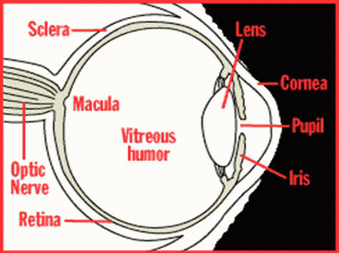
Anatomy of the Eye
The Cornea
This is a very thin, clear structure, allowing
light to get into the eye and focusing it
through the pupil onto the lens. In some
people it is irregular or cone-shaped ,
notfocusing light correctly. Glasses or surgery
such as LASIX may be required to correct this.
The Lens
The lens of the eye is just behind the pupil. It
collects light and focuses it on the macular
part of the retina. It is normally a clear
crystalline structure. In cataract formation, it
begins to get cloudy and does not let the light
through.
The Retina
The retina, which senses light rays, is a thin
lining on the back of the eye. The macula,
which is affected in macular degeneration, is a
very small spot in the center of the retina. It is
here that the ability to read, identify small and
distant objects and other tasks requiring good
vision is located.
Diabetic retinopathy and macular degeneration
are the most common conditions affecting the
retina and macula.
The Optic Nerve
The optic nerve transmits the image from the
retina to the brain where the image is
processed. If the nerve is not normal or is
damaged, the image may be distorted,
incomplete or even absent. This is usually
called optic neuritis or neuropathy, and may be
from a variety of conditions. Glaucoma , by
raising the pressure inside the eye, affects the
small nerve fibers in the eye. Multiple sclerosis
affects the nerve after it leaves the eye, and
stroke may stop the processing of the image
by the brain.

FOLEY, AL
251-721-1160

Copyright © CSVRLowVision.org. All Rights Reserved. Website hosted by North Mobile Internet Services, Inc.
LOW VISION AND THE EYE
WHAT IS LOW VISION?
Vision less than normal, not corrected by medical treatment or glasses, interfering with normal activities such
as reading and driving.
CAUSES OF LOW VISION AND BLINDNESS:
•
Macular degeneration *(see link below for self-diagnosis)
•
Diabetes
•
Glaucoma
•
Other
*link for self-diagnosis of AMD, supplied by the Macular Degeneration Foundation. Click link to see video.
http://www.mdsupport.org/amdtest.html
WHAT CAN BE DONE TO HELP?
From space-age technology to simple adaptive changes in the home, many things can be done to improve the
function and independence of those
with low vision or blindness. At our offices we have a full selection of non-optical, optical and electronic
devices, both high and low tech.
STANDARD EYE CARE:
Our clinics are not intended to provide routine eye care. We function only as a rehabilitative service.
WHO WILL YOU SEE?
You will be seen first by one of our doctors. A thorough evaluation of your vision will be performed. This will
include functional tests not ordinarily done by your Eye Doctor. This evaluation will show what training, devices
and instruction will help. The doctor may have you return to see the Occupational Therapist(OT). The OT will
provide training in the changes and adaptations needed for comfortable living with low vision or blindness.
Instruction in the many ways to achieve independence and the use of devices will be taught by the OT. You
may be referred to other teachers, agencies, or resources if they can help in any way.
Anatomy of the Eye
The Cornea
This is a very thin, clear structure, allowing light to
get into the eye and focusing it through the pupil onto
the lens. In some people it is irregular or cone-
shaped , notfocusing light correctly. Glasses or
surgery such as LASIX may be required to correct
this.
The Lens
The lens of the eye is just behind the pupil. It collects
light and focuses it on the macular part of the retina.
It is normally a clear crystalline structure. In cataract
formation, it begins to get cloudy and does not let the
light through.
The Retina
The retina, which senses light rays, is a thin lining on the back of the eye. The macula, which is affected in
macular degeneration, is a very small spot in the center of the retina. It is here that the ability to read,
identify small and distant objects and other tasks requiring good vision is located.
Diabetic retinopathy and macular degeneration are the most common conditions affecting the retina and
macula.
The Optic Nerve
The optic nerve transmits the image from the retina to the brain where the image is processed. If the nerve is
not normal or is damaged, the image may be distorted, incomplete or even absent. This is usually called optic
neuritis or neuropathy, and may be from a variety of conditions. Glaucoma , by raising the pressure inside the
eye, affects the small nerve fibers in the eye. Multiple sclerosis affects the nerve after it leaves the eye, and
stroke may stop the processing of the image by the brain.

251-476-4744 in Mobile 251-928-2888 in Daphne 251-721-1160 in Foley


